Pathology imaging systems for the autopsy suite and grossing workstation have evolved well beyond standalone cameras and manual photo logs, providing integrated documentation, annotation, and specimen tracking capabilities that support both clinical quality management and medicolegal chain-of-custody requirements. ARES Scientific sources imaging systems from Mopec and SPOT Imaging — two manufacturers whose products address the full range of documentation needs within the pathology and mortuary equipment workflow, from high-resolution gross specimen photography to bulk barcode-based sample tracking. These systems serve hospital pathology departments, surgical pathology labs, medical examiner offices, and academic anatomy programs, supporting the documentation standards required by CAP accreditation, CLIA compliance, and forensic case management. When integrated with a facility's laboratory information system (LIS), they eliminate manual transcription steps and reduce the risk of specimen identification errors that represent one of the most consequential failure modes in pathology laboratory operations. ARES Scientific's coverage of anatomy, morgue, and pathology facilities extends across the complete suite of postmortem and surgical pathology infrastructure.
Types of Pathology Imaging Systems
Autopsy Imaging Systems
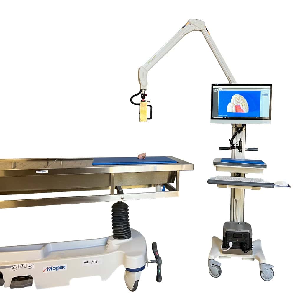
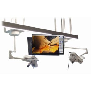
Autopsy imaging systems are designed to provide structured, reproducible photographic documentation throughout the postmortem examination — from external body surface findings to organ section photography following dissection. The Mopec PathCam platform, available in HD and MICRON configurations and deployable on a mobile station, integrates directly with grossing workstations to eliminate the need for a separate photography setup. The integrated design keeps the camera and lighting in a fixed, reproducible position relative to the specimen, which is important for maintaining consistent image quality across different cases and operators. In the forensic context, image consistency and traceability are essential: photographs taken during an autopsy may be introduced as evidence in legal proceedings, and any break in the documentation chain — including inconsistent framing, lighting, or file naming — can be challenged. These systems support autopsy table workflows by keeping photographic documentation within arm's reach of the prosector rather than requiring a separate photography station setup for each case.
Gross Imaging Systems
Gross imaging systems focus specifically on high-resolution photography of surgical and autopsy specimen sections during the grossing process. The Mopec PathCam Gross Imaging System delivers 20 MP still image capture with 10x optical zoom and 12x digital zoom, autofocus, and a maximum field of view of 28″×36″ for full-organ captures, narrowing to a minimum field of approximately 0.185″×1″ for fine-detail specimen close-ups. Video output supports 1920×1080p at up to 60 fps, as well as 1280×720p, providing flexibility for facilities that incorporate video review into their gross examination workflow. The practical implication of this resolution and zoom range is that a single camera system can document a full bowel resection specimen at arm's length and then zoom into a suspicious mucosal lesion without repositioning the specimen or changing equipment. This range of capability makes the system viable across surgical pathology, autopsy, and educational settings. The gross imaging subcategory provides direct access to this product family.
Sample Tracking and Documentation Systems
Sample tracking systems address a distinct but closely related documentation challenge: ensuring that every specimen container, cassette, or block in the laboratory is correctly identified and associated with the right case throughout the processing workflow. The SPOT Imaging PathTracker Bulk Barcode Reader reads up to 150 barcodes in 25 seconds and uploads the data automatically to the laboratory information system, providing visual confirmation of each scanned specimen batch through an intuitive graphical interface. Manual barcode scanning of individual specimens is a known source of transcription errors and workflow bottlenecks in high-volume labs; bulk scanning eliminates both problems simultaneously. The system's direct integration with the LIS creates an auditable electronic record of specimen receipt, processing, and handoff that supports CAP accreditation documentation requirements. The sample tracking and documentation subcategory covers this product line in detail.
Key Features and Technology in Pathology Imaging Systems
Integrated Workstation Design and Annotation Capability
One of the most significant operational advantages of purpose-built pathology imaging systems over general-purpose cameras is the integration of the imaging hardware with the grossing workstation environment. The Mopec PathCam systems are designed to fit seamlessly into grossing workstations, eliminating the need to move specimens to a separate photography area or adjust ad-hoc camera setups between cases. Integrated digital annotation tools allow the pathologist or pathology assistant to label findings, add measurements, and notate images directly within the imaging interface, without exporting files to a separate annotation program. This capability is important for CAP and ISO 15189 compliance, both of which require that pathology documentation be traceable, legible, and associated with the correct specimen record. When combined with LIS connectivity, annotations become part of the permanent case record rather than a separate image archive that must be manually linked. Facilities evaluating these systems should confirm LIS compatibility before specifying, as integration requirements vary by platform. These systems are designed to work alongside grossing stations as a coordinated documentation infrastructure.
Image Resolution, Zoom Range, and Field of View
The resolution and optical performance specifications of a pathology imaging system determine which specimen types and examination tasks the system can support reliably. At 20 MP with 10x optical zoom and a field of view spanning from 28″×36″ down to sub-inch detail, the PathCam Gross Imaging System covers the full range from large organ specimens — kidneys, liver lobes, brain sections — to the fine margin detail that surgical pathology requires for resection specimens. The 12x digital zoom extends reach further without requiring physical repositioning, though optical zoom is preferable for diagnostic imaging because it maintains image quality without the pixel interpolation artifacts introduced by digital zoom. For video output, 1920×1080p at 60 fps is sufficient for real-time gross examination review and remote teaching applications. Facilities that include gross examination in resident or student education should prioritize video quality and streaming capability alongside still-image resolution when evaluating imaging systems.
Barcode Scanning Speed, LIS Integration, and Chain-of-Custody Documentation
The operational value of a bulk barcode reader like the PathTracker is best understood against the baseline it replaces: a technician scanning individual specimen containers one at a time, manually verifying each against a paper log, and periodically reconciling the physical count against the LIS. At 150 barcodes per 25 seconds with automatic LIS upload and visual confirmation, the PathTracker replaces this process with a single workflow step that is simultaneously faster, more accurate, and automatically documented. In medicolegal and forensic pathology contexts, this level of documentation is particularly important: chain-of-custody for every specimen must be demonstrable, and gaps in the electronic record can create evidentiary problems in legal proceedings. Hospital pathology departments subject to CAP inspection also benefit from the auditable specimen tracking log the system creates. The SPOT Imaging manufacturer page provides additional technical background on the PathTracker platform and its integration architecture.
Applications of Pathology Imaging Systems Across Facility Types
Hospital Surgical Pathology Departments
Surgical pathology departments in hospital settings process resection specimens, biopsies, and organ specimens from operating room cases daily, generating a continuous documentation requirement that scales directly with surgical volume. A dedicated gross imaging system positioned at the grossing station allows the pathology assistant or pathologist to photograph each specimen in a consistent, reproducible format — with controlled lighting, a fixed focal distance, and integrated annotation — without interrupting the grossing workflow to retrieve or set up a camera. CAP accreditation requires that photographic documentation practices be defined and consistently applied; a purpose-built system with standardized positioning and LIS connectivity is far easier to validate and audit than a workflow relying on variable personal devices or shared cameras. Specimen tracking through bulk barcode reading reduces the risk of specimen mislabeling or misassociation with cases, which the College of American Pathologists identifies as a significant quality and safety concern in surgical pathology. Hospital procurement teams evaluating imaging systems should assess the volume of daily cases against the system's throughput capacity and LIS integration compatibility before finalizing a specification.
Medical Examiner and Forensic Pathology Offices
Medical examiner offices have documentation requirements that differ from clinical pathology in both purpose and legal consequence. Photographs taken during an autopsy in a medicolegal case become part of the official case record and may be used in criminal or civil legal proceedings, meaning image traceability, date-time stamping, and chain-of-custody documentation are not optional — they are foundational requirements of forensic practice. An integrated autopsy imaging system that records each image with metadata linking it to the case file and examiner eliminates the ambiguity that arises when photographs are taken with a personal device and transferred manually to a case record. The PathCam platform on a mobile station is well suited to medical examiner environments because it can be repositioned between multiple autopsy bays as needed, providing consistent documentation capability across the suite without fixed installation at every table. These offices typically benefit from a coordinated approach that combines imaging systems with autopsy carts and autopsy tables designed to support multi-modal examination workflows.
Academic Anatomy and Pathology Education Programs
University anatomy programs, medical schools, and pathology residency programs use gross imaging systems primarily as teaching tools — capturing specimen photographs for case presentations, image libraries, and remote learning content. The video output capabilities of the PathCam system, including 1080p at 60 fps, make it suitable for live demonstration of gross examination technique to groups of students who cannot all stand at the table simultaneously. High-resolution still images support the development of educational atlases and case archives that residents and students can review outside of the grossing room. Academic anatomy programs that handle willed body donations may also benefit from the documentation consistency that an integrated imaging system provides, ensuring that anatomical findings are recorded systematically across a teaching cohort. Coordination with related equipment — including dissection tables and specimen handling infrastructure — supports a well-documented, educationally structured anatomy suite.
Selecting Pathology Imaging Systems for Your Laboratory
Matching System Capability to Case Volume and Specimen Type
The primary selection criteria for a pathology imaging system should reflect the facility's actual case mix and daily throughput. A surgical pathology department processing 50 or more gross specimens per day has different requirements than a small community hospital morgue handling occasional clinical autopsies. High-volume labs benefit most from systems with rapid autofocus, wide field-of-view flexibility, and seamless LIS upload — capabilities that reduce per-case documentation time at scale. Lower-volume facilities with complex specimen types — unusual anatomical presentations, forensic cases, or teaching material — may prioritize image resolution, annotation depth, and video streaming quality over throughput speed. Gross imaging and autopsy imaging systems address overlapping but distinct use cases, and facilities that perform both surgical pathology grossing and postmortem examinations may find that specifying one system for each workflow — rather than a single system deployed across both — produces better results per case type.
LIS Integration and IT Infrastructure Requirements
LIS compatibility is the most consequential technical requirement to verify before specifying any pathology imaging or specimen tracking system. Bulk barcode readers and integrated imaging platforms both create structured data that must map correctly to the existing case management and specimen tracking fields in the facility's LIS. Incompatible data formats, unsupported upload protocols, or gaps in field mapping can render the system's automation benefits inaccessible and require manual workarounds that negate the efficiency gains. Facilities should engage their LIS vendor and IT department in the evaluation process before selecting an imaging or tracking system, confirming that the required integration is supported and that the implementation timeline is feasible. Mobile system configurations — such as the PathCam on a mobile station — also require evaluation of power access, cable management, and cart mobility within the specific floor plan of the pathology suite or autopsy room.
Vendor Support, Software Updates, and Long-Term Serviceability
Pathology imaging systems combine hardware — camera optics, mounting hardware, lighting — with software that manages annotation, file naming, LIS communication, and user interface functions. The software component introduces a long-term serviceability consideration that does not apply to purely mechanical pathology equipment: operating system compatibility, software licensing, and vendor update support all affect the useful life of the system. Facilities should evaluate the manufacturer's track record for software support, the availability of training resources for new staff, and the terms of any service or support agreements before finalizing a purchase. The ARES Scientific pathology and mortuary equipment catalog provides access to both Mopec and SPOT Imaging product lines, allowing facilities to evaluate gross imaging and specimen tracking systems from a single source alongside the broader suite of autopsy and grossing infrastructure.
ARES Scientific supports hospital pathology departments, medical examiner offices, and academic anatomy programs in identifying and specifying imaging systems from Mopec and SPOT Imaging, alongside the full range of pathology and mortuary equipment needed to outfit a complete postmortem and surgical pathology suite.