Pathology is a cornerstone of medical diagnosis and has undergone a significant transformation over the centuries. From the early days of rudimentary microscopes to today’s sophisticated digital platforms, the field has continually adapted to incorporate more advanced technologies. This evolution not only enhances the accuracy of diagnoses, but also streamlines the processes that underpin the modern healthcare industry. This article explores the historical development of pathology, the impact of transitioning to digital methods, and introduces a revolutionary tool: the digital grossing station equipped with a camera that interfaces seamlessly with digital pathology programs.
The Evolution of Pathology: A Historical Perspective
As a medical discipline, pathology has roots deeply embedded in the annals of medical history, starting from the natural human curiosity about the causes of disease and death. Initially intertwined with general medical practice, the formal discipline of pathology did not emerge until much later. The earliest recorded observations date back to ancient civilizations like Egypt, where the Edwin Smith Papyrus and the Ebers Papyrus (circa 1600 BCE) documented cases of tumors and infections, hinting at the beginnings of pathological anatomy. However, these documents were not systematic studies of disease, but rather collections of observations without a cohesive structure or a scientific methodology.
The intellectual leap in pathology came with the Greeks, particularly from Hippocrates and later, the Alexandrians like Herophilos and Erasistratos, who began to correlate clinical practice with post-mortem examinations. Although the practice was sporadic and not widely accepted due to cultural and religious taboos, these early endeavors laid the groundwork for a more structured approach to understanding human diseases. Herophilos, in particular, is noted for his attempts to correlate structure with function, a foundational concept in pathology.
The true scientific methodology in pathology began to take shape during the Renaissance, a period marked by a revived interest in human anatomy and dissections. This period saw figures like Antonio Benivieni of Florence, who systematically recorded case histories and autopsy findings, set the stage for pathology as a distinct scientific discipline. The Renaissance also brought advancements in the preservation and examination of tissues, further contributing to the anatomical understanding of disease.
By the 19th century, pathology was firmly established as a scientific discipline, largely thanks to the work of doctors like Giovanni Morgagni, who is often called the father of modern pathology. Morgagni’s work, “De Sedibus et Causis Morborum per Anatomen Indagatis” (“The Seats and Causes of Diseases Investigated by Anatomy”), published in 1761, systematically linked clinical symptoms with post-mortem findings, laying the foundation for pathological anatomy. This period also saw the development of tools and techniques such as microscopy, which allowed for the detailed study of tissues and cells, further cementing pathology’s role as a critical scientific field in medicine.
The Impact of Microscopy on Pathology and the Transition to Digital Pathology

The Advent of Microscopy and Its Influence on Pathology
The development of the microscope in the late 16th century by inventors such as Zacharias Janssen revolutionized the biological sciences, profoundly impacting the field of pathology. With the ability to magnify small objects, early microscopes allowed scientists like Robert Hooke and Antonie van Leeuwenhoek to observe cells and microorganisms for the first time. This capability laid the foundational understanding of tissue structure and disease pathology. As microscopes improved, so did the ability to diagnose and understand diseases at a cellular level, transitioning pathology from a macroscopic to a microscopic science.
Enhancements in Microscopic Techniques and Pathological Practices
Throughout the 19th and 20th centuries, enhancements in staining techniques and the development of electron microscopes provided pathologists with detailed views of the cellular arrangements and structures within tissues. These advancements enabled the identification and categorization of numerous pathological conditions based on cellular morphology and tissue architecture. Diseases could now be diagnosed with greater accuracy, facilitating targeted treatment strategies that significantly improved patient outcomes.
Challenges in Traditional Pathology
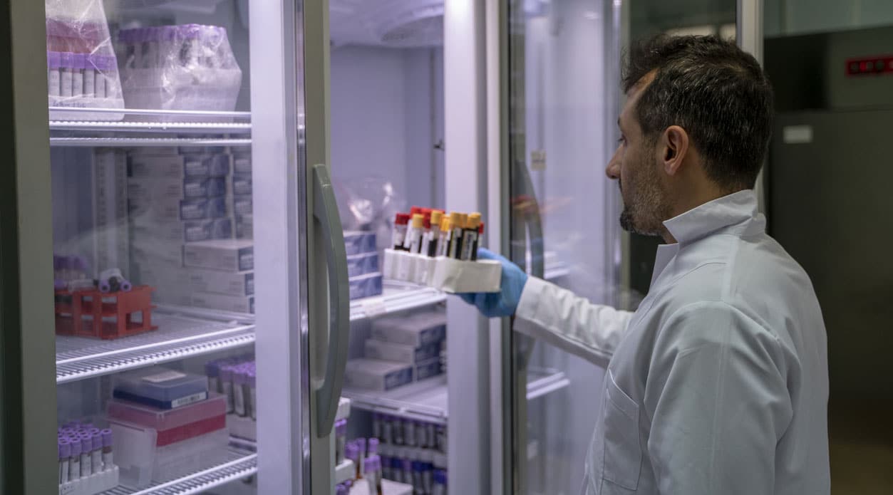
Despite its advancements, traditional microscopy-based pathology faced limitations. The physical nature of slide preparation, storage, and examination limited the speed and efficiency of pathological assessments. Physical slides were prone to degradation, and their analysis required the physical presence of skilled pathologists, which could delay diagnoses, particularly in remote or resource-limited settings.
Introduction of Digital Pathology: A Paradigm Shift

The advent of digital pathology marked a significant shift in how pathological assessments are conducted. Digital pathology involves scanning traditional glass slides to create high-resolution digital images that can be viewed, shared, and analyzed using computers. This transition not only preserves the integrity of samples but also enhances accessibility and collaboration among pathologists worldwide, overcoming geographical limitations and accelerating diagnostic processes.
The Role of Epic LIS and Epic Beaker in Digital Pathology: Enhancing Diagnostic Accuracy and Efficiency
Epic Systems Corporation, a leader in healthcare technology, provides Epic LIS and Epic Beaker, which are integral tools in the digital pathology landscape. Epic Beaker, in particular, streamlines the workflow from specimen collection to diagnosis. It supports an integrated approach where digital images and traditional laboratory data coexist, enhancing diagnostic accuracy. The system enables pathologists to access comprehensive clinical and radiological patient data, facilitating informed and precise diagnostic decisions.
Enhancing Diagnostic Precision with Advanced Data Management
Epic Beaker improves diagnostic precision through advanced data management. It supports tissue-based workflows crucial for digital pathology, integrating seamlessly with other Epic applications. This integration centralizes patient information, reducing errors and improving diagnostic quality. Managing large volumes of data and combining it with digital imaging technologies allows pathologists to review and share digital slides, enhancing collaborative diagnosis efficiently.
Impact of Digital Pathology on Modern Healthcare
Digital pathology, complemented by artificial intelligence and machine learning, offers tools that significantly improve diagnostic accuracy and speed. AI algorithms help analyze digital slides to detect anomalies, aiding pathologists in delivering faster and more accurate diagnoses. Furthermore, the capability for remote consultations in digital pathology enables specialists to contribute globally, enhancing the scope and quality of medical diagnostics.
Streamlining Operations and Improving Turnaround Times
Epic Beaker significantly boosts laboratory efficiency by automating tasks and standardizing workflows. This automation speeds up the diagnostic process and facilitates quicker patient management decisions, which is critical in modern healthcare settings.
Future Perspectives and Continued Evolution
As digital pathology continues to evolve, its integration with technologies like AI and machine learning is expected to enhance diagnostic precision and efficiency further. The ongoing development of sophisticated AI models and the standardization of digital pathology practices will solidify its role as an indispensable tool in modern medicine. This evolution is set to improve patient outcomes through faster, more accurate diagnoses and revolutionize educational and research activities in pathology.
Advancements in Pathology Labs: Mopec Maestro Encore and PathCam Systems
Revolutionizing Pathology with Advanced Grossing Stations
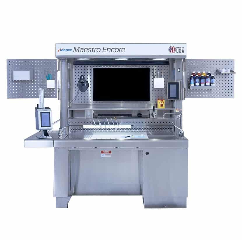
In the dynamic field of pathology, where precision and efficiency are paramount, the integration of advanced technology like the Mopec Maestro Encore Grossing Station significantly enhances laboratory operations. This state-of-the-art station embodies the latest advancements in laboratory technology, seamlessly blending the meticulous preparation of specimens with the sophisticated capabilities of digital imaging and analysis.
Mopec Maestro Encore: A Comprehensive Grossing Solution
The Mopec Maestro Encore Grossing Station is distinguished by its incorporation of the PathCam Gross Imaging System. This integration marks a significant evolution from traditional microscopy, enabling pathologists to transition smoothly between gross examination and microscopic analysis. The built-in PathCam allows for the immediate capture and digital documentation of specimen images at the grossing station, which expedites the entire diagnostic process from specimen receipt to analysis, ensuring rapid and accurate results crucial for effective patient treatment, especially in a critical diagnosis like cancer.
Seamless Electronic Health Record Integration and Regulatory Compliance
One of the most notable features of the Mopec Maestro Encore is its compatibility with leading electronic health record systems such as Epic and Epic Beaker. This functionality allows for the effortless capture, annotation, and direct upload of images into a patient’s health record, streamlining data management and ensuring that detailed, precise information accompanies every diagnostic step. Additionally, the system’s design and functionality have received approval from rigorous bodies, including the United States Department of Veterans Affairs, highlighting its reliability and adherence to high standards of medical care.
![]()
ARES: Your Pathology Equipment Specialists
When it comes to equipping modern pathology labs with the latest in diagnostic technology, ARES stands out as your dedicated specialist. ARES provides comprehensive solutions tailored to the evolving needs of pathology, emphasizing the integration of innovative systems like the Mopec Maestro Encore and the PathCam Gross Imaging System. These tools are crucial for labs aiming to enhance their diagnostic capabilities and efficiency, ensuring that they are well-equipped to meet the demands of modern healthcare.
The Role of PathCam in Enhancing Digital Pathology
The PathCam Gross Imaging System further exemplifies technological innovation in pathology. This system is pivotal for the field’s transition towards fully digital operations, offering high-resolution imaging capabilities that are integral for accurate diagnostics. By facilitating the capture and annotation of detailed images during the gross examination phase, PathCam not only enhances the accuracy of diagnostics, but also supports educational and collaborative efforts across medical communities.
Future Outlook: Integrating Cutting-Edge Pathology Solutions
As pathology continues to evolve, the integration of advanced technologies like the Mopec Maestro Encore with digital pathology systems is expected to play a central role. These technologies improve diagnostic accuracy and streamline processes and ensure compliance with health record standards. Looking ahead, the continued adoption and integration of such technologies will bridge the gap between traditional microscopy and digital pathology, leading to improved patient outcomes and more efficient healthcare systems.
By partnering with ARES for their pathology equipment needs, labs can leverage the expertise and cutting-edge solutions necessary to advance their diagnostic capabilities. This partnership ensures that labs are equipped with the best tools to meet the increasing demands of speed, accuracy, and compliance in healthcare, propelling them into the future of digital pathology. For more information on how ARES can enhance your laboratory’s capabilities, please contact us today.